Neuronal Programs
 Glial Programs
 Circuit Function
 Human Translation
How metabolic state rewires brain circuits
Metabolic stress-induced transcriptional programs driving cortical circuit vulnerability in neurodegeneration.
Explore the research
Insulin resistance alters cortical inhibitory neurons and microglia to exacerbate Alzheimer’s phenotypes
Metabolic stress worsens memory in Alzheimer’s mice by reprogramming glia and inhibitory neurons and destabilizing cortical circuits.
Molecular Neurodegeneration, 2026.
Metabolic vulnerability in neurodegeneration
Systemic metabolic stress, a state where the body struggles to obtain or utilize the nutrients it needs, underlies many chronic health conditions and diseases, including obesity, type 2 diabetes, and cardiovascular disease. This condition can also impair cognition and is increasingly recognized as a significant contributor to neurodegenerative disease onset and progression. This is especially true for Alzheimer’s disease, which is now considered a metabolic stress disorder. My research centers on how this metabolic stress drives cognitive decline in Alzheimer’s disease by altering transcriptional programs that govern brain circuit stability. I am particularly interested in the differential engagement of these programs across distinct neuronal subtypes and glial populations, which contribute to early circuit dysfunction in neurodegenerative diseases.
Using an integrated, multi-level approach, I determine how systemic metabolic stress disrupts brain function at different scales. At the mechanistic level, I identify cell type-specific transcriptional programs in neurons and glia, determining how they regulate intrinsic cell physiology, immune signaling, and synaptic function. I then explore how these programs translate to changes in circuit dynamics and brain cognition, prioritizing those relevant to human disease to identify translationally significant therapeutic targets. This framework, applied in a clinically relevant Alzheimer’s disease mouse model, aims to define early mechanisms of cognitive decline and reveal general principles that extend beyond any single neurological disorder.

Explore each research area →
My approach: from molecular mechanisms to translational relevance
Mouse genetics
I have established a clinically relevant Alzheimer’s disease mouse model, combined with metabolic stress paradigms and conditional genetics, to interrogate how stress-induced transcriptional programs regulate cellular physiology, from single-cell properties to circuit function.
Omics
With the power of tools such as single-nucleus sequencing, ChIP-seq, and IP-MS, I can pinpoint metabolic stress programs and the genetic and molecular mechanisms that underlie them.
Microscopy
I use confocal and STED microscopy to validate omics hits and link them to changes in glial and neuronal morphology, synapses, and brain anatomy.
Cellular electrophysiology
Using ex vivo whole-cell patch clamp, I test how metabolic stress alters intrinsic cellular properties and synaptic function, linking transcriptional programs to their functional consequences.
Circuit tools
Using cell-type-specific Cre driver lines and Cre-dependent chemogenetic tools (DREADDs), I selectively silence or activate defined neuronal populations to map their circuit connections and test their contributions to circuit function.
Human validation
I leverage public human cortical datasets (ROSMAP, SEA-AD, ACT) to test whether the transcriptional programs identified in mice are present in human disease and to identify therapeutic targets.
Metabolic control of neuronal excitability
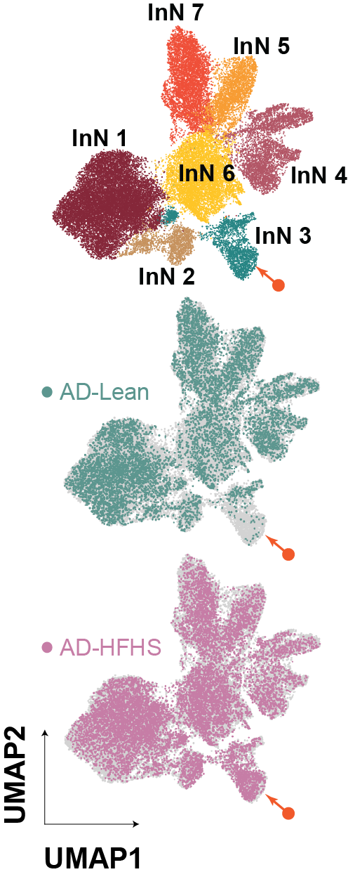
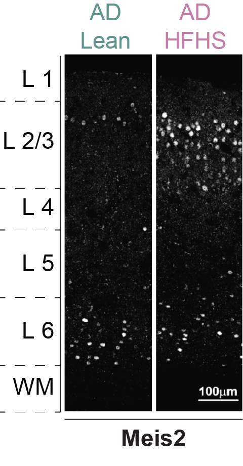
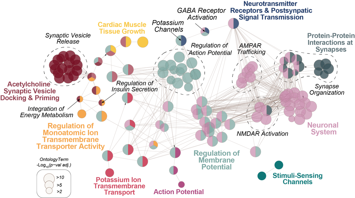
Alzheimer’s disease is a devastating age-related metabolic stress disorder. Most cases are sporadic, driven by environmental factors like diet and lifestyle rather than a single mutation, a pattern shared with other metabolic disorders such as autism spectrum disorder and Parkinson’s disease. Diabetes, or even chronic hyperglycemia, often emerges years before AD diagnosis, and this metabolic stress can alter gene regulation to the detriment of neuronal and circuit function. A key feature of AD is cortical hyperexcitability, arising from a disrupted excitatory-inhibitory balance that impairs learning and memory. I have identified a metabolic stress-responsive transcriptional program in cortical inhibitory neurons of the AD brain, marked by signatures of altered excitability. These programs may act as a transcriptional node linking metabolic impairment to inhibitory circuit dysfunction, leading to disinhibition and hyperexcitable cortical networks. I’m now using genetic tools and cellular electrophysiology to define what these programs regulate and how they alter the excitability of vulnerable neurons under metabolic stress. This work will lay the foundation for investigating whether distinct metabolic stress-responsive programs govern other cell types and circuit populations.
Metabolic control of glial function
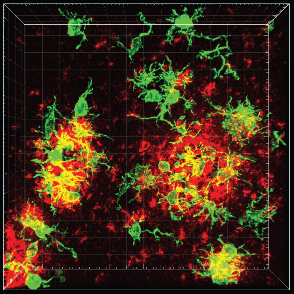
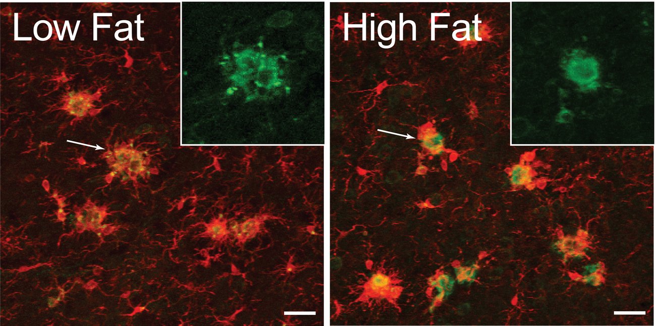
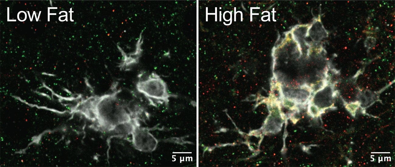
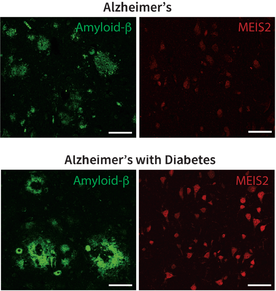
Microglia are the brain’s resident immune cells, and in Alzheimer’s disease, their activation state can shift from adaptive responses to a persistent inflammatory state. Building on prior evidence of glial-mediated synapse loss in aged AD mice, my work examines how metabolic dysfunction alters the disease-associated microglial activation state. In disease-associated microglia, I observed remodeling of microglial morphology and membrane-associated proteins linked to synapse organization and signaling. These changes were part of a distinct, coordinated pan-glial transcriptional program, which I termed the metabolic impairment in neurodegeneration (MinD) state and found to extend across microglia, astrocytes, and oligodendrocytes. This program is associated with selective inhibitory synapse loss, suggesting a link between glial remodeling and circuit vulnerability. Future studies will investigate whether the glial MinD state represents a neuroprotective adaptive response, a maladaptive driver of pathology, or a context-dependent state that shifts over the course of disease progression.
Metabolic control of circuit function
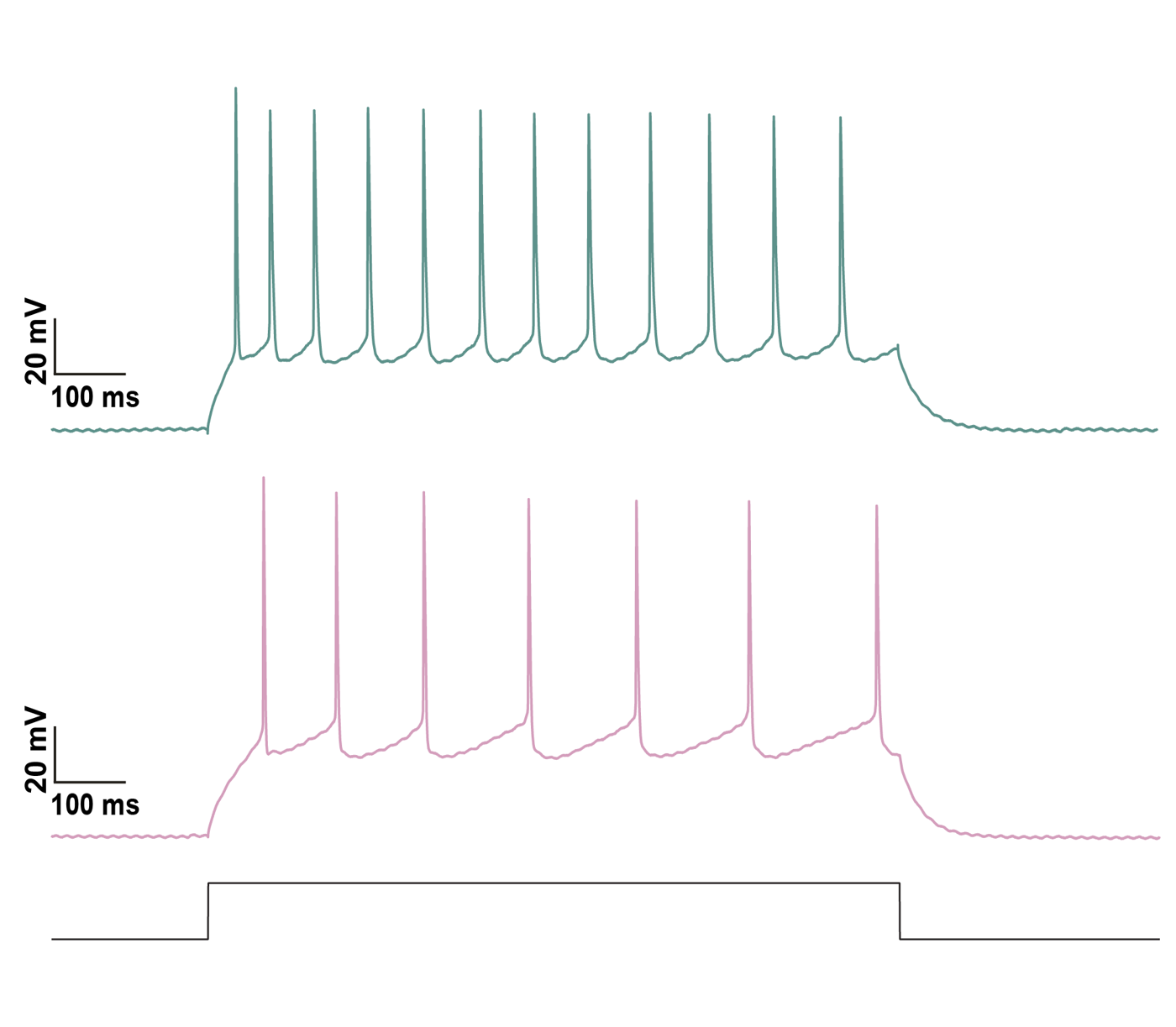
Cortical circuits depend on coordinated activity across neurons and glia. Yet it remains unclear how metabolic stress engages distinct molecular programs across cell types to alter cellular function and drive circuit-level dysfunction. I found that diet-induced insulin resistance induces cell-type-specific transcriptional programs in cortical inhibitory neurons and glial cells in the AD mice. These programs are accompanied by signatures of altered inhibitory neuronal excitability, a selective loss of inhibitory synapses, and exacerbated cognitive deficits. Together, these findings suggest that metabolic stress engages transcriptional programs in both inhibitory neurons and glia that may predispose cortical networks to dysfunction. By combining mouse genetics with ex vivo and in vivo electrophysiology and circuit mapping, I test how these coordinated programs shape cortical circuit dynamics during disease progression.
Metabolic control of human disease
Metabolic dysfunction, including impaired glucose regulation and insulin resistance, is an early predictor of Alzheimer’s disease (AD) risk and cognitive decline. However, it remains unclear whether cell-type-specific transcriptional programs identified in mouse models are conserved in the human AD cortex or associated with clinical outcomes. To address this, I integrate postmortem human cortical tissue with large-scale public datasets (ROSMAP, SEA-AD, ACT) to test whether these programs are enriched in individuals with metabolic comorbidities. This approach bridges mechanistic insights from mouse models with human disease and aims to identify conserved transcriptional signatures that may serve as therapeutic targets.

I’m a neuroscientist and Kavli Postdoctoral Research Fellow at Yale University, with a background in biomedical sciences and translational research. I study how systemic metabolic stress alters brain function in Alzheimer’s disease.
Why I study how extrinsic cues converge to drive intrinsic cellular programs that govern neuronal function?
This question has been the driving force behind my career, beginning with my first research experience. As an undergraduate, I studied how metabolic stress alters gene expression to drive bacterial virulence, where I first became interested in stress-responsive gene regulation and the idea that environmental cues can alter cellular behavior. I then worked in a spinal cord injury (SCI) research group, where I developed viral vectors and cell transplantation protocols for axonal regeneration and studied the mechanisms underlying spontaneous regeneration. This work raised a key question: why do neurons differ in their intrinsic capacity to respond to injury?
In my Master’s research at the MPI for Brain Research, I developed a quantitative imaging approach to map newly synthesized proteins in neuronal compartments, showing that neuronal molecular architecture is functionally compartmentalized and responsive to localized extrinsic signals. As a doctoral student with Prof. Amparo Acker-Palmer at Goethe University Frankfurt, I defined how VEGF from glial, neuronal, and vascular sources signals through compartment-specific receptor trafficking to regulate hippocampal circuit development. Together, these studies established how the cellular environment instructs neuronal structure and function through spatially defined signaling.
Now, I use transcriptomic approaches to study how metabolic stress affects different cell types in Alzheimer’s pathophysiology. These experiences have converged on my current effort: to understand how metabolic stress reprograms the intrinsic physiological state of neurons and glia in Alzheimer’s disease, and, ultimately, other neurodegenerative diseases.
Why is human translational research important to me?
Before my Ph.D. studies, I worked for several years with Dr. Armin Blesch as a research technician in spinal cord injury (SCI) research, first at UCSD and later at the University of Heidelberg SCI Clinic in Heidelberg, Germany. Our group focused on targeted gene therapy-based approaches to promote axonal regeneration and circuit formation after SCI, while also targeting key aspects of the injury site, including neurodegeneration, glial scar formation, and inflammation. The move to Heidelberg offered an immensely rewarding opportunity to transition from a traditional academic environment to a more interdisciplinary, patient-oriented setting. At the SCI clinic in Heidelberg, I worked with doctors, basic research scientists, engineers developing robotics and prosthetic limbs, and physiotherapists. Together, we worked not only to treat but also to understand the full clinical picture of our patients, with the goal of improving clinical treatments and patient mobility.
This interdisciplinary experience left me with two profound lessons that have continued to shape my scientific career. First, with input from patients who had accepted the loss of walking ability and become more focused on quality of life, my former boss completely changed his research focus from neuronal regeneration to pain and bladder control. Second, the gap between our understanding of neuronal function across scales, from genes to physiology to circuits, and our ability to manipulate the regenerative properties of neurons remains too large for direct implementation into patient therapies. Neither of these lessons would have resonated with me as deeply without the direct interactions and input I received from the clinic’s patients. Because of this, I was driven to pursue my master’s and Ph.D. in neuroscience, aiming to build a scientific career with a specific focus on human translational research.
How I invest in the potential of people, in science, and in the communities that connect them?
I am proud to have helped build neuroscience outreach and training programs that support the next generation of scientists. As a PhD student in Frankfurt, I co-founded the Deutsche Neurowissenschaften Olympiade e.V. (DNO), the German national branch of the International Brain Bee, a neuroscience competition that inspires high school students to pursue careers in neuroscience. Together with my co-founders, I helped expand the program to four German cities (Berlin, Bonn, Heidelberg, and Frankfurt) and led a network of more than 60 postdocs and PhD students who engaged approximately 200 students annually. At the Frankfurt location, I led a 16-member team, trained scientists in science communication, and secured a five-year educational partnership with the Hertie Foundation. I also helped launch Brain Bee programs in the Netherlands and Argentina, broadening my perspective on how neuroscience education can connect students and scientists across countries and training environments.
I also worked to address structural gaps in academic career development. I created a teaching training program that provided postdocs at non-university research institutions, including the Max Planck Institutes, with a pathway to earn habilitation teaching credits. I organized workshops connecting PhD students with pharmaceutical and technology companies and led career development events attended by 200–300 participants. Across these initiatives, I secured more than €250,000 to support neuroscience education and cross-institutional collaboration at local, national, and international levels. I am proud that many of the programs I helped initiate are still running today, led by dedicated people who continue to sustain and expand them. As a postdoc at Yale, I have continued my commitment to neuroscience outreach and community building. In addition to mentoring and training students, I am coaching local community leaders as they develop mentoring programs for children. I am also working on my first neuroscience book for children, which follows a young girl as she explores and understands the world around her through science.
Papers & preprints
Organized by research area. *co-first / first author
Alzheimer’s disease & neurodegeneration
Transcriptomics · metabolic stress · brain circuits.
Spinal cord injury & axon regeneration
AAV-based gene therapy · iPSCs · biomaterials.
Protein dynamics in synapse and circuit assembly
Live cell imaging · hippocampal circuits · neurovascular signaling · protein trafficking.
Microbial & Bacterial Physiology & Metabolism
Bacterial stress response · gene regulation.
Honors, talks & highlights
A running log of talks, honors, papers, and milestones I’m proud of.
Research support
My research draws on and is supported by the following consortia, data resources, and funding:
Professional development
Programs I’ve taken part in — shared here so others can find and apply to them too:
Coursework & training
Selected postdoctoral coursework and training I’ve completed:
Beyond the bench